Repiration notes
RESPIRATION
L1. STRUCTURE AND FUNCTION
L1. STRUCTURE AND FUNCTION
Respiration Diagram Key
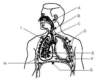
A. Pharynx: - common passage for respiratory and digestive systems above the epiglotis
B. Larynx: - Structure that contains the vocal cords, voice box (just below epiglotis)
C. Trachea: - the windpipe (cartilagenous ridges)
I. Bronchi: - The two major divisions of the trachea leading to the lungs
- Divides further in each lung into smaller passages called bronchioles
Bronchioles: - The smaller brances of tubes in the lungs
- each bronchiole ends in an air pocket sac called an alveoli
E. Alveoli: - Saclike structures that are the air sacs of a lung
- makes up the lungs
G. Diaphragm: - a sheet of muscle that separates the chest cavity from the abdominal cavity.
D. Ribs: - bones hinged to the vertebral column and sternum which, with muscle, define the top and sides of the chest cavity.
H. Pleural Membranes (4): - membranes that enclose the lungs
- Outer pleural membrane sticks closely to the walls of the chest and the diaphragm
- Inner pleural membrane is stuck to the lungs
- The two lie very close to each other
- Pressure between the two is less than outside air pressure or lungs collapse.
- Stick lungs to chest cavity walls
- lubrication
F. Thoracic Cavity: - Sealed off chest, where lungs are.
- Ribs form top and sides, diaphram forms bottom
- used in inspiration and expiration
L2. CILLIA AND MUCUS
Cillia line the tubes of the respiratory tract. The tubes also produce mucus, which traps bacteria and dust particles. The cillia sweep the mucus upward, cleaning the resp. tubes.
L3. ALVEOLI
- 700,000 alveoli in human lung, 100x surface of skin
- thin walled sacs covered with a capillary network
- CO2 and O2 can diffuse directly through the walls
- site of O2 / CO2 exchange
L4. & L5. INHALATION AND EXHALATION
BREATHING
- The taking in of air into the lungs.
in - inspiration
out - expiration
The lungs lie in a sealed off cavity - chest or thoracic cavity
- the ribs make up the top and sides, the diaphram makes up the bottom, the pleural membranes seal it.
L6. ROLE OF CO2, H+, AND MEDULLA OBLONGATA
- the urge to breathe is brought about primarily by CO2 / H+ ions
- monitored in the blood by the medula oblongata - resp. centre in brain
- low O2 is monitored by chemo receptors in the carotid artery (neck). Low O2 will cause the rate and depth of breathing to increase.
- High CO2 stimulates breathing centre which stimulates diaphram and rib muscles to contract. You breathe in.
- when lungs are filled, stretch receptos on the alveoli send messages to the breathing centre, which shut down signals to the diaphram and rib muscles. They relax and you breathe out.
L7. 7 L8. GAS EXCHANGE AND TRANSPORT
External (at Alveoli)
- the CO2, O2 exchange in the alveoli
- relies on diffusion
High CO2-----> Low CO2
(blood) (air)
Low O2<------- High O2
(blood) (air)
CO2 is carried in blood in three ways
1) as bicarbonate (aprox. 70%)
A. Pharynx: - common passage for respiratory and digestive systems above the epiglotis
B. Larynx: - Structure that contains the vocal cords, voice box (just below epiglotis)
C. Trachea: - the windpipe (cartilagenous ridges)
I. Bronchi: - The two major divisions of the trachea leading to the lungs
- Divides further in each lung into smaller passages called bronchioles
Bronchioles: - The smaller brances of tubes in the lungs
- each bronchiole ends in an air pocket sac called an alveoli
E. Alveoli: - Saclike structures that are the air sacs of a lung
- makes up the lungs
G. Diaphragm: - a sheet of muscle that separates the chest cavity from the abdominal cavity.
D. Ribs: - bones hinged to the vertebral column and sternum which, with muscle, define the top and sides of the chest cavity.
H. Pleural Membranes (4): - membranes that enclose the lungs
- Outer pleural membrane sticks closely to the walls of the chest and the diaphragm
- Inner pleural membrane is stuck to the lungs
- The two lie very close to each other
- Pressure between the two is less than outside air pressure or lungs collapse.
- Stick lungs to chest cavity walls
- lubrication
F. Thoracic Cavity: - Sealed off chest, where lungs are.
- Ribs form top and sides, diaphram forms bottom
- used in inspiration and expiration
L2. CILLIA AND MUCUS
Cillia line the tubes of the respiratory tract. The tubes also produce mucus, which traps bacteria and dust particles. The cillia sweep the mucus upward, cleaning the resp. tubes.
L3. ALVEOLI
- 700,000 alveoli in human lung, 100x surface of skin
- thin walled sacs covered with a capillary network
- CO2 and O2 can diffuse directly through the walls
- site of O2 / CO2 exchange
L4. & L5. INHALATION AND EXHALATION
BREATHING
- The taking in of air into the lungs.
in - inspiration
out - expiration
The lungs lie in a sealed off cavity - chest or thoracic cavity
- the ribs make up the top and sides, the diaphram makes up the bottom, the pleural membranes seal it.
- Inspiration
- diaphram contracts (lowers)
- rib muscles contract (pull up and out)
This expands the thoracic cavity which causes a low pressure, air is "sucked" in.
- Expiration
- diaphram relaxes (raises)
- ribs relax
Thoracic cavity relaxes (gets smaller) and air is forced out.
L6. ROLE OF CO2, H+, AND MEDULLA OBLONGATA
- the urge to breathe is brought about primarily by CO2 / H+ ions
- monitored in the blood by the medula oblongata - resp. centre in brain
- low O2 is monitored by chemo receptors in the carotid artery (neck). Low O2 will cause the rate and depth of breathing to increase.
- High CO2 stimulates breathing centre which stimulates diaphram and rib muscles to contract. You breathe in.
- when lungs are filled, stretch receptos on the alveoli send messages to the breathing centre, which shut down signals to the diaphram and rib muscles. They relax and you breathe out.
L7. 7 L8. GAS EXCHANGE AND TRANSPORT
External (at Alveoli)
- the CO2, O2 exchange in the alveoli
- relies on diffusion
High CO2-----> Low CO2
(blood) (air)
Low O2<------- High O2
(blood) (air)
CO2 is carried in blood in three ways
1) as bicarbonate (aprox. 70%)
- The enzyme Carbonic anhydrase catalyzes these reactions in the RBC
2) -some of the remaining CO2 is carried as dissolved CO2 dissolved directly in the plasma (CO2 is reasonably soluble in H2O.
3) - the rest of the CO2 is carried by hemoglobin directly as HbCO2
Oxygen is carried in the blood only one way
O2 is carried in blood directly on hemoglobin
Hb + O2----->HbO2
- hemoglobin accepts O2 easier at cooler, neutral pH in lungs and gives it up at warmer, more acid (lower pH) environment of the tissues .
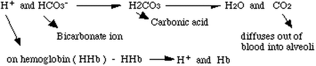
2. Internal (Tissues)
- CO2 - O2 exchange in body tissue
a) Oxygen
HbO------> 2Hb + O2
O2 diffuses into cells from blood
b) CO2 (the three ways again)
-70% of CO2 is carried as bicorbonate ion (HCO3-) dissolved in plasma
2) -some of the remaining CO2 is carried as dissolved CO2 dissolved directly in the plasma (CO2 is reasonably soluble in H2O.
3) - the rest of the CO2 is carried by hemoglobin directly as HbCO2
Oxygen is carried in the blood only one way
O2 is carried in blood directly on hemoglobin
Hb + O2----->HbO2
- hemoglobin accepts O2 easier at cooler, neutral pH in lungs and gives it up at warmer, more acid (lower pH) environment of the tissues .
2. Internal (Tissues)
- CO2 - O2 exchange in body tissue
a) Oxygen
HbO------> 2Hb + O2
O2 diffuses into cells from blood
b) CO2 (the three ways again)
-70% of CO2 is carried as bicorbonate ion (HCO3-) dissolved in plasma
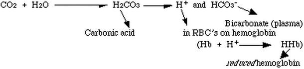
- The enzyme Carbonic anhydrase catalyzes these reactions in the RBC
- remaining 30% of CO2 is carried as:
i) HbCO2 (carbamino hemoglobin )
ii) or CO2 dissolved directly in blood
Gas Exchange
Gasses (CO2, O2) easily exchange due to the process of diffusion
Internal
i) body tissue - O2 is low (always used up)
- CO2 is high (being produced)
ii) blood - O2 is high (HbO2)
- CO2 is low
External
i) alveoli - O2 is high (20% of air)
- CO2 is low (.5% of air)
blood - O2 is low (diffused out in body tissue)
- CO2 is high (produced in body tissue, carried as HCO3-
